OPTICAL INSTRUMENT

IN-SITU RAMAN + ELECTROCHEMISTRY
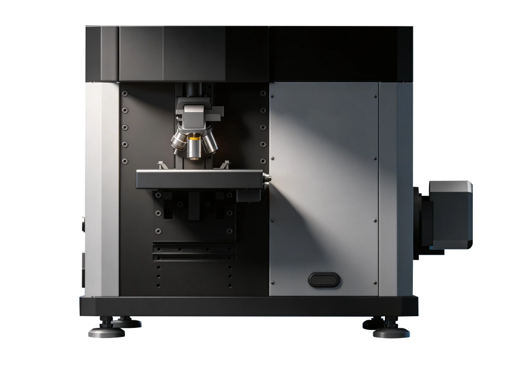
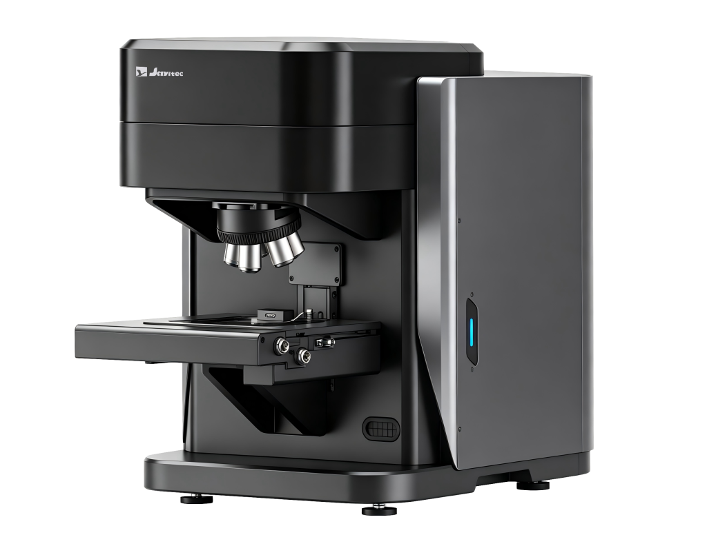
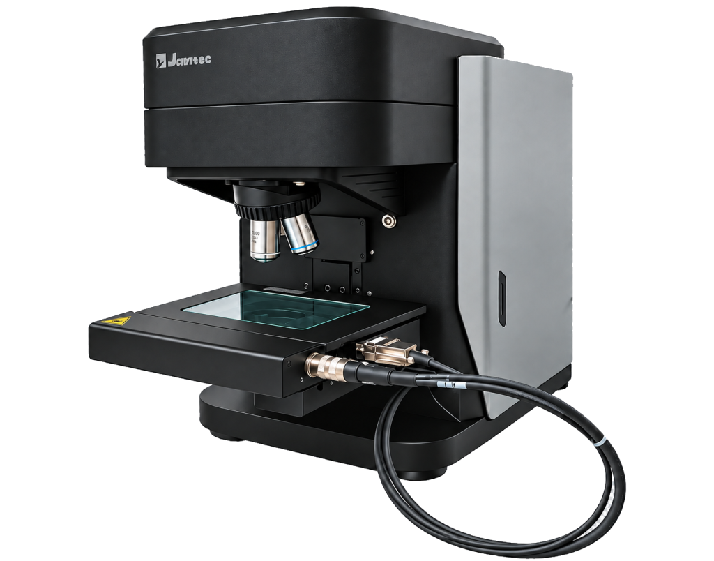
EC-Raman seamlessly integrates three traditionally independent systems—microscopic imaging and scanning, Raman spectroscopy, and electrochemical analysis—into one unified platform.
By integrating the optical, electronic, and control systems within a single instrument, EC-Raman eliminates the complexity of conventional setups that rely on multiple instruments, cables, and software platforms. Its unified architecture overcomes key limitations of externally coupled systems, including incompatible communication protocols, unpredictable transmission delays, and fragmented data acquisition, enabling synchronized and reliable electrochemical Raman measurements.
A patented magnetic electrochemical cell further simplifies operation, enabling tool-free connection and sample loading within seconds. This significantly reduces setup time and improves experimental efficiency and reproducibility.
Flexible Excitation for Diverse Samples
Choose the optimal excitation wavelength for different sample characteristics. With broad spectral coverage from low wavenumbers to the near-infrared, EC-Raman combines optimized lasers, detectors, filters, and gratings to deliver high-quality Raman spectra with excellent signal-to-noise performance.
Deep-Cooled, Low-Noise Detector
Deep cooling effectively suppresses thermal noise, enabling sensitive detection of weak Raman signals. Even during rapid acquisition, EC-Raman delivers clean, stable, and reproducible spectra with reduced background interference and baseline drift, ensuring reliable measurements with high signal quality.
Upright Reflective Microscope
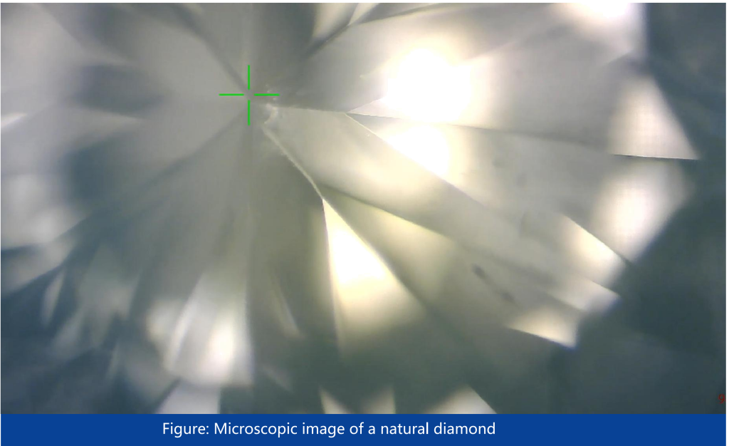
High-brightness LED illumination, bright-field optics, and a high-resolution camera provide clear and detailed sample visualization. A 5-position objective turret with 10×, 50×, and 100× objectives supports flexible observation from rapid large-area sample locating to high-magnification analysis.
High-Definition Chemical Imaging
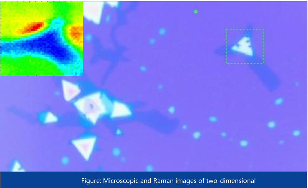
Supports point, line, and area Raman mapping, precisely correlating spectral information with spatial locations. High-resolution chemical images reveal the composition and structural distribution of samples, enabling intuitive visualization of chemical and material heterogeneity.
Three Flexible Stage Options
Three stage configurations—domestic open-loop, imported open-loop, and imported closed-loop—are available to meet different requirements, from routine Raman mapping to precise micro-area positioning. The system delivers positioning accuracy up to ±1 μm and Raman imaging resolution <1 μm, enabling precise and reproducible spatial analysis.
Particle Positioning Accuracy <2 μm
Automatically identifies micron-scale particles and precisely positions each target at the laser focus using the high-precision motorized stage, enabling automated Raman acquisition with positioning accuracy <2 μm.
Reliable Electrochemical Control for Transient Raman Analysis
A wide potential range and high current output support diverse electrochemical applications. ~100 ns hardware-triggered synchronization precisely coordinates electrochemical control with Raman acquisition, ensuring each Raman spectrum accurately corresponds to its electrochemical state.
Transient Electrochemical Raman Technology
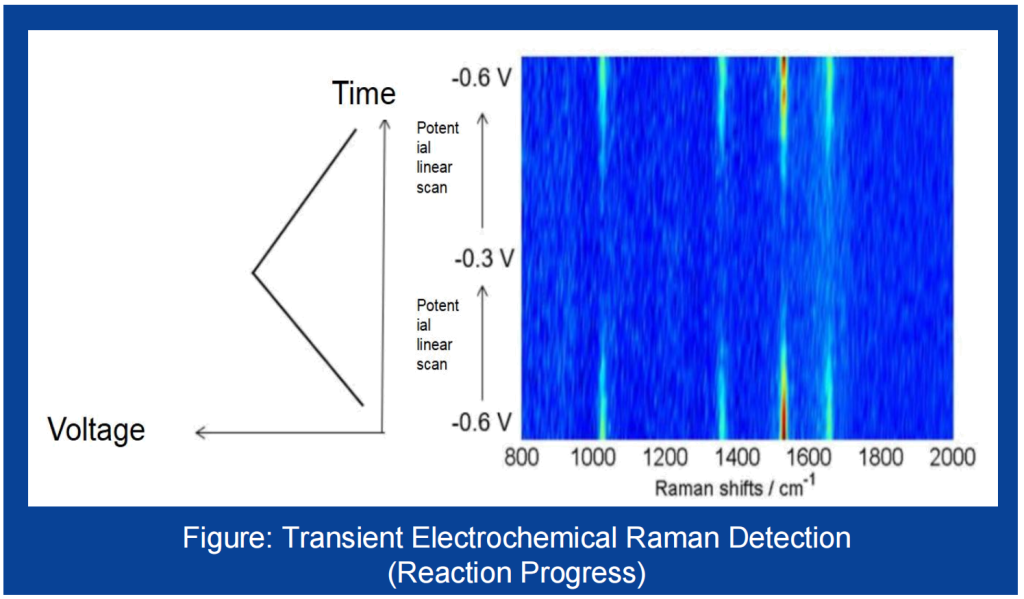
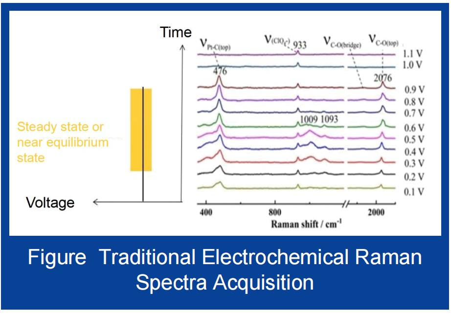
Electrochemical reactions often involve short-lived intermediates that form and disappear on millisecond or even microsecond timescales. Conventional electrochemical Raman measurements typically follow a “stabilize the potential, then acquire the spectrum” workflow, providing mainly averaged information under steady-state or near-equilibrium conditions. Like taking a static photograph, this approach can miss critical transient processes during a reaction.
In 2015, our chief scientist pioneered Transient Electrochemical Raman spectroscopy, first introducing the methodology in the Journal of the American Chemical Society (JACS). By synchronizing Raman acquisition with continuous potential scanning or rapid potential steps, the technology enables real-time tracking of molecular species at electrode interfaces—directly revealing the formation, transformation, and disappearance of reaction intermediates.
This provides time-resolved molecular-level insights into electrocatalysis, battery interfaces, corrosion mechanisms, and other dynamic electrochemical processes. If conventional electrochemical Raman takes a “snapshot,” Transient Electrochemical Raman records a “video”—revealing not only where a reaction starts and ends, but how it actually happens.
Implementation Approach
Transient Electrochemical Raman places extremely high demands on the synchronization of the instrumentation: electrochemical excitation and Raman acquisition must be precisely aligned on the same time axis.
EC-Raman addresses this challenge at the hardware level,
Hardware Trigger Synchronization
The main control unit coordinates the electrochemical module and Raman module. As the electrochemical excitation signal changes, the controller simultaneously sends a hardware trigger to initiate Raman acquisition, achieving a synchronization delay of approximately 100 ns. This near-zero-delay synchronization provides a reliable temporal foundation for capturing transient Raman signals.
TECHNICAL SPECIFICATIONS
| Product type | Integrated electrochemical Raman microscope |
| Excitation wavelengths | 532 nm / 633 nm / 785 nm options |
| Spectral resolution | <4 cm−1 |
| Spectral Repeatability | ≤ 0.2 cm⁻¹ |
| High-Precision Motorized Stage | XYZ Travel Range: 75 × 50 × 50 mm Minimum Step Size: ≤ 50 nm Repeatability: ≤ 1 μm XY Positioning Accuracy: ≤ ±1 μm Raman Imaging Resolution: ≤ 1 μm |
| Microscope | Upright reflective microscope; bright-field LED; high-sensitivity camera |
| Objectives | 5-position turret with 10×, 50×, and 100× objectives 10× Objective:NA: 0.30, WD: 8.5 mm 50× Objective:NA: 0.5, WD: 10.6 mm 100× Objective:NA: 0.90, WD: 1.0 mm |
| Spectral Range | 170–3900 cm⁻¹ @ 532 nm 170–3500 cm⁻¹ @ 633 nm 170–3400 cm⁻¹ @ 785 nm |
| Positioning performance | Stage accuracy up to ±1 µm; particle positioning <2 µm; Raman imaging <1 µm |
| Electrochemical techniques | Full range of experimental modes: CV, LSV, SCV, TAFEL, CA, CC, DPV, NPV, DNPV, SWV, ACV, SHACV, FTACV, i-t, DPA, DDPA, TPA, IPAD, BE, HMV, SSF, PITT, IMP, AC Impedance (Potentiostatic Control), IMPT, IMPE, CP, CPCR, ISTEP, GTT, PSA, ECN, OCPT, Constant-Potential Electrolysis, RDE Control (0–10 V Output), AC Impedance Data Simulation and Fitting Program |
| Potential Ranges | ±10 V, ±1 V, ±100 mV, ±10 mV |
| Applied Potential Accuracy | 0.1% FS ± 1 mV |
| Current Control Range | ±250 mA |
| Current Ranges | 2 nA–250 mA, 10 ranges in total |
| Applied Current Accuracy | 0.1% FS |
| Electrochemical cell | Magnetic three-electrode cell |
| Dimensions | 340 × 630 × 460 mm |
| Software | EX-Raman |
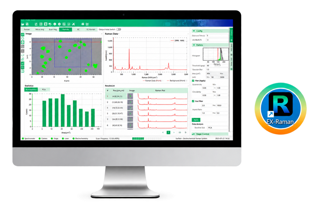
Unified Control & Data Analysis
EX-Raman integrates Raman acquisition, microscopy, electrochemical control, mapping, particle analysis, and data processing into one unified platform. Point, line, and area Raman mapping precisely correlates spectral data with spatial positions for intuitive chemical imaging and analysis.
Automated & Synchronized Analysis
Automated particle detection enables rapid identification, positioning, and Raman analysis of micron-scale particles. Synchronized Raman and electrochemical data acquisition enables real-time tracking of molecular evolution and reaction intermediates at electrode interfaces.
APPLICATIONS
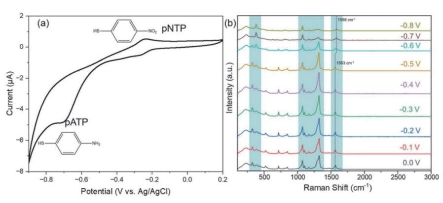
Reaction pathways and molecular evolution are monitored in real time through potential contral and in-situ Raman spectroscopy.
The electrochemical CV curve shows an increasing reduction current with decreasingpotential In-situ Raman spectra reveal that as the potential shifts from 0 to -0.5 V, thecharacteristic nitro group peak of pNTP (-1330 cm 1) gradually decreases, while theamino group peak of pATP (-1593 cm increases. The Raman spectral evolution closelymatches the electrochemical reduction region in the CV curve, directly revealing themolecular conversion of pNTP to pATP and ena bling real-time tracking of interfacialreaction processes.
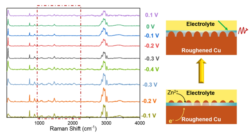
Using potential-controlled in-situ Surface-Enhanced Raman Spectroscopy (SERS), with roughened copper (Roughened Cu) serving as both the SERS substrate and working electrode, the system enables real-time monitoring of interfacial evolution in zinc-based electrolyte systems over the charging/discharging potential range (0.1 V to −0.3 V vs. reference electrode). The results reveal the formation of electrolyte reduction products and provide molecular-level insights into battery interfacial reaction mechanisms.
RESOURCES
RELATED PRODUCTS
OPTICAL INSTRUMENT
OPTICAL INSTRUMENT
OPTICAL INSTRUMENT