OPTICAL INSTRUMENT

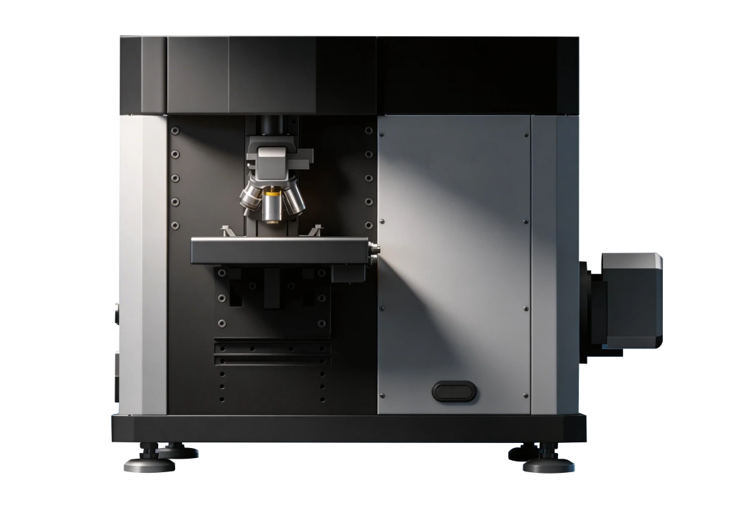
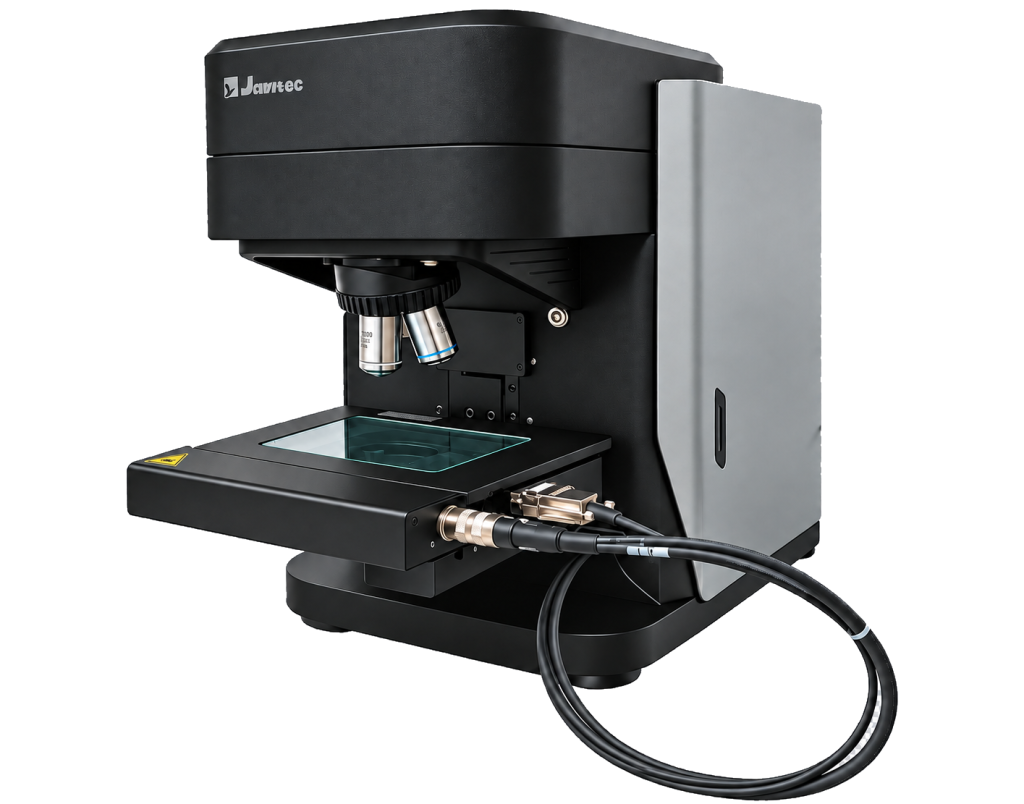
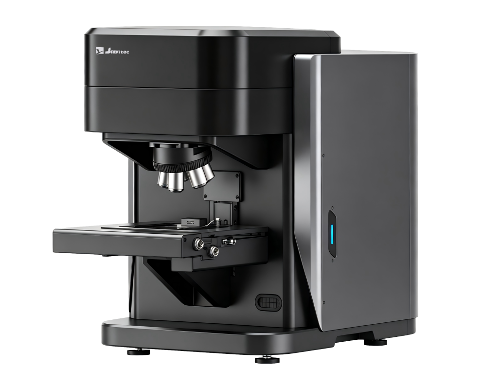
Intelligent Automated Raman Microscope
Particle Recognition, Auto Positioning, Raman Mapping,Batch Analysis
OVERVIEW
Smart Raman builds on Xiamen University’s decades of academic excellence and engineering expertise in Raman spectroscopy. Xiamen University has continuously pioneered advances in surface-enhanced Raman spectroscopy (SERS), tip-enhanced Raman spectroscopy (TERS), and advanced Raman instrumentation.
Smart Raman embodies this technological heritage, combining cutting-edge Raman spectroscopy with intelligent instrument design. Backed by decades of expertise in Raman microscope development, SmartRaman sets a new benchmark for intelligent Raman microscopy in spatial resolution, spectral resolution, detection speed, and sensitivity.
The system is designed to meet advanced research needs across materials science, biomedical research, forensic science and security, environmental monitoring, and other fields.
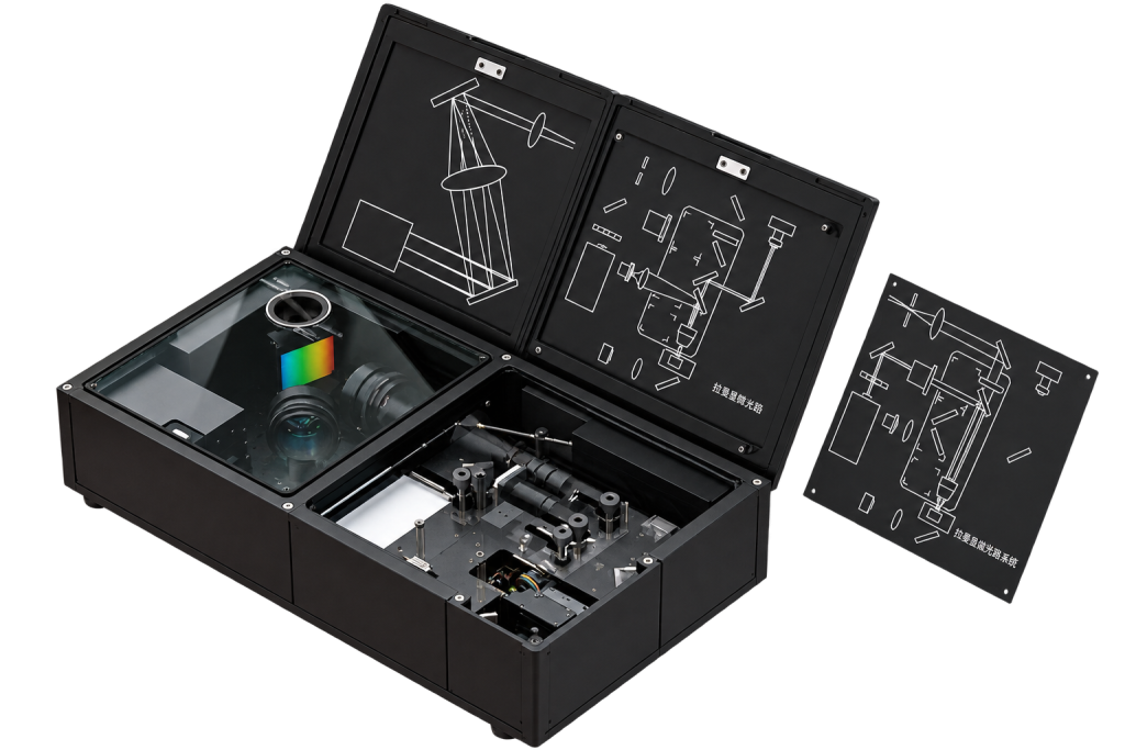
The system overcomes the limitations of conventional Raman microscopes—such as external optical paths, complex fiber connections, and bulky configurations—through a highly integrated architecture that combines the Raman microscope module and high-precision spectrometer into a single compact platform. By eliminating external fiber coupling, the system minimizes space requirements and can be readily installed inside a glovebox, providing greater flexibility for in-situ experiments and laboratory configurations.

High-Resolution Microscopic Imaging
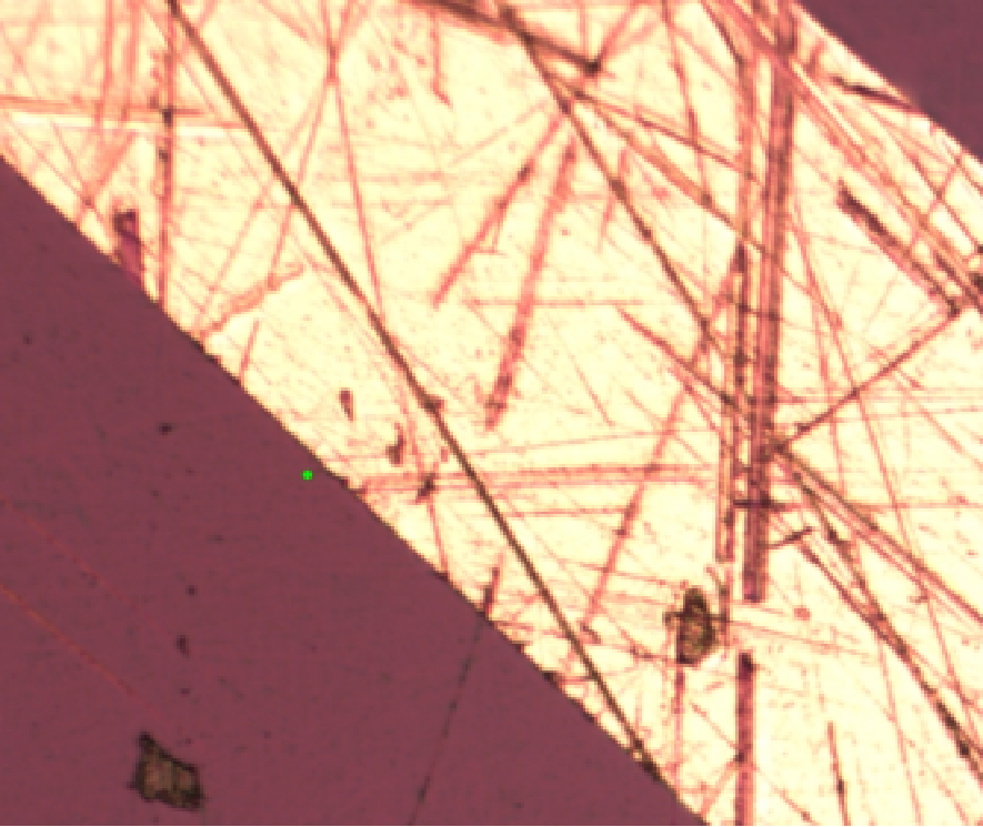
Equipped with an 8-megapixel color camera and LED bright-field illumination, the system delivers clear, detailed visualization of sample surfaces. From rapid target localization to precise micro-region identification, it enables efficient and intuitive microscopic observation throughout the Raman analysis workflow.
Key features
01
02
03
Advanced Raman Software Platform
From spectral acquisition to data analysis, the highly integrated software provides a seamless workflow within a single interface. Particle recognition is included as standard, with optional Raman–electrochemistry integration for expanded research capabilities.
Integrates Raman spectroscopy, microscopic imaging, Raman mapping, particle detection, and data analysis in one platform, enabling centralized control and real-time data synchronization throughout the workflow.

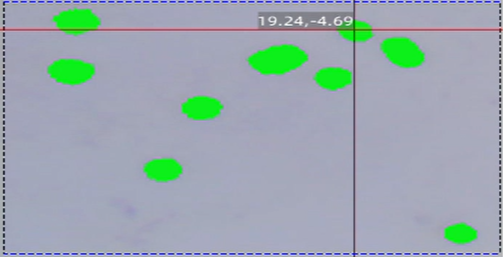
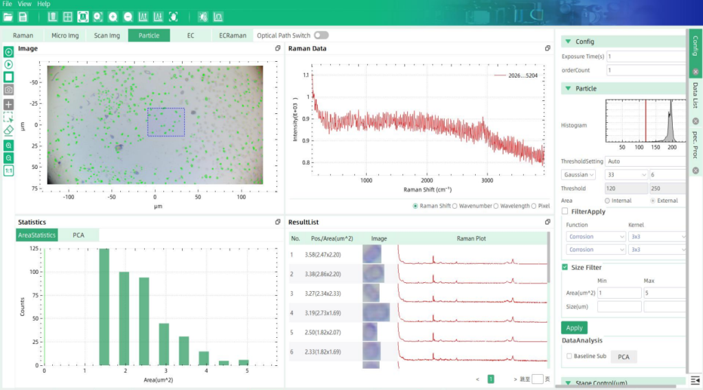
Automatically identifies particles using adaptive thresholding, morphological filtering, and size filtering, enabling one-click particle detection and batch Raman acquisition.
Provides comprehensive data management and traceability, ensuring organized, reliable, and reproducible research data.
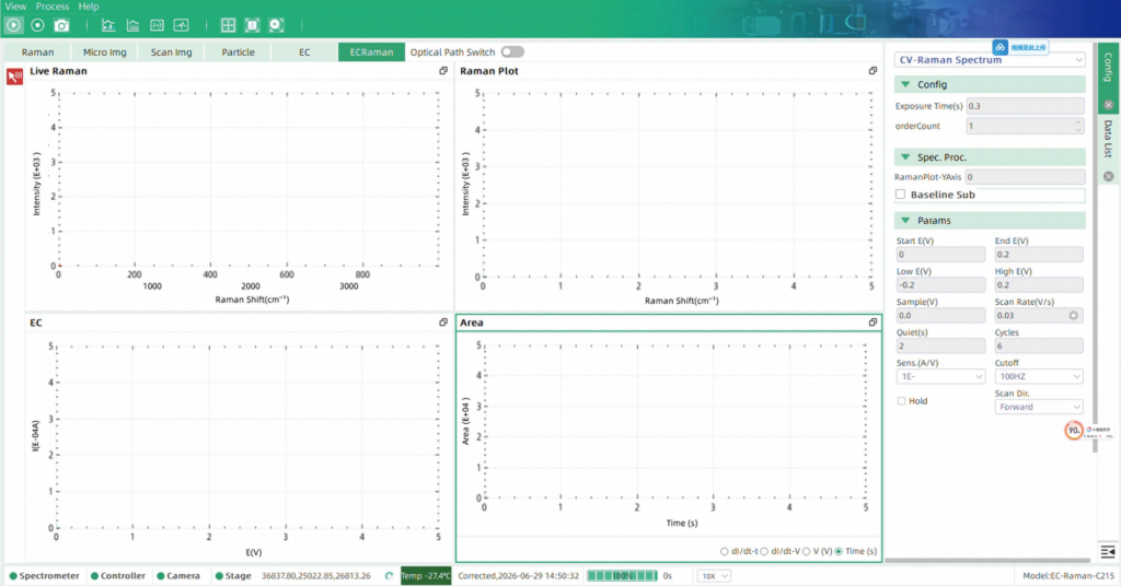
The optional Raman-electrochemical coupling module enables synchronized acquisition and real-timevisualization of Raman and electrochemical signals within a unified interface. It allows dynamic monitoring ofmolecular structural evolution and the formation and disappearance of species at the electrode interface.
Application Scenarios
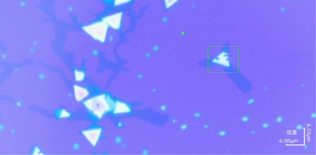
Smart Raman enables rapid, non-destructive Raman imaging of 2D materials with high spatial resolution and optical stability, revealing interlayer coupling, structural uniformity, and defect distribution for in-depth materials characterization.
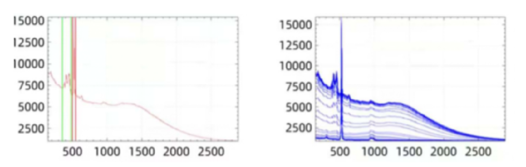
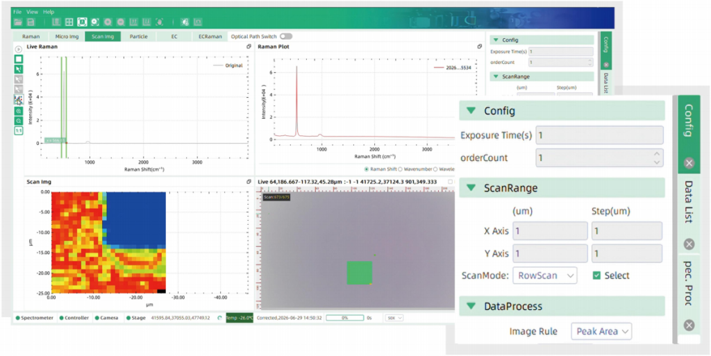
Real-time Raman spectra of a single pixel and spectra from all pixels of MoS2
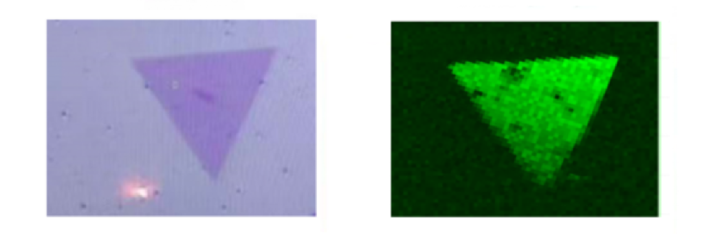
Microscopic image and Raman mapping image of MoS2
The system simultaneously acquires Raman signals from the in-plane vibrational mode Eg (387 cm1)and the out-of-plane vibrational mode Aig (405 cm1). Based on the spatial intensity distributiondifferences between these two signals, it can reconstruct high-resolution Raman imaging in real time.
Each pixel in the resulting image reflects differences in color intensity corresponding to the material’s spatial distribution, structural uniformity, and potential defect regions.
For rapid and non-destructive layer number identification, the peak separation (A) between A.g and Egmodes is calculated. When A falls precisely within the range of 17-20 cm1, the region can be identifiedas a monolayer structure, enabling fast and reliable thickness determination.
Microplastic particles as small as 1 um are traditionally identified through time-consuming manualmicroscope screening and individual spectral acquisition, with limited efficiency and reproducibility. SmartRaman integrates an intelligent particle recognition algorithm that enables automatic localization ofmicroplastic particles and batch acquisition of Raman spectra for each particle. The software simutaneouslygenerates statistical reports, including particle count, size distribution, and composition ratios, allowing clear visualization of microplastic distribution and source analysis
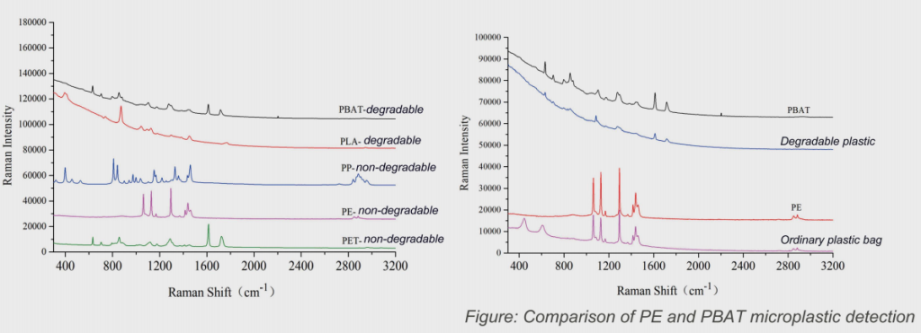
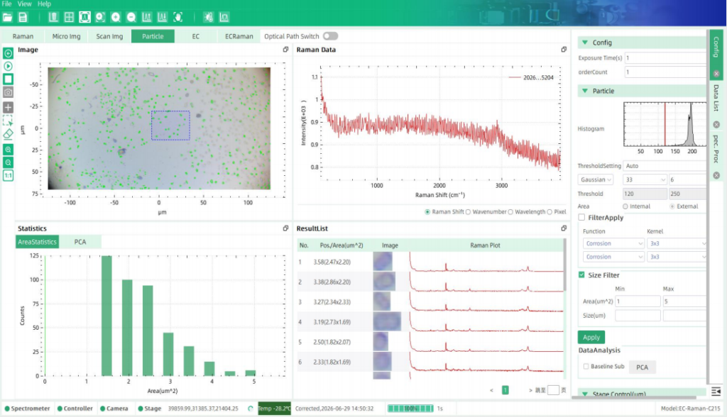
Batch microplastic detection using particle recognition
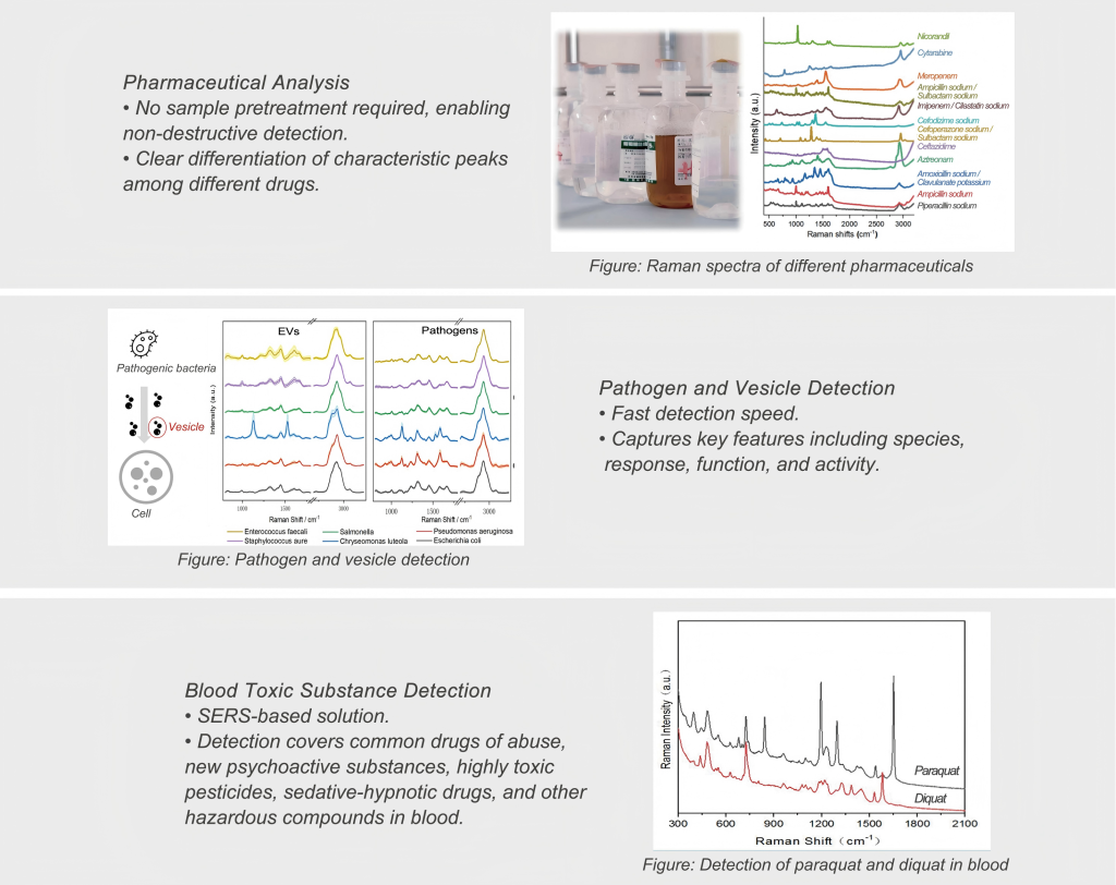
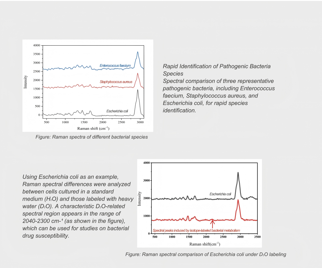
Without the need for complex fluorescence staining or antibody labeling, the intrinsic “molecularfingerprint” capability of Raman spectroscopy enables direct qualitative analysis of tissue sections,single cells, and even pathogenic microorganisms. This is crucial for preserving the native state ofsamples and avoiding interference from labeling agents.Combined with the SERS technology developed by the Xiamen University, Smart Raman enableshighly sensitive detection of trace substances in complex biological matrices such as blood. Thisprovides a new spectroscopic perspective for early diagnosis, pathological mechanism research,
and drug delivery monitoring.
RESOURCES
RELATED PRODUCTS
OPTICAL INSTRUMENT
OPTICAL INSTRUMENT
OPTICAL INSTRUMENT